Infected Nonunion Tibia Case
Infected Nonunion: Tibia Case 1
Presenting
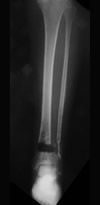
Initial x-ray and photograph of an infected tibial nonunion in a 66 year old woman. The patient had been treated at an outside facility by other physicians using a plate and screws. The plate had been removed and approximately one inch of bone had been removed by another surgeon before the patient was referred in to Dr. Brinker's office for reconstruction.
Treatment in Progress
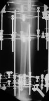
This x-ray taken during treatment shows Ilizarov bone transport in progress with bone grafting at the nonunion site (near the ankle).
Result After Treatment by Dr. Brinker
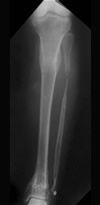
This final x-ray shows a healed distal tibial nonunion and restoration of normal bone length. The patient has returned to his pre-injury activity.
Infected Nonunion: Tibia Case 2
Presenting
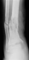
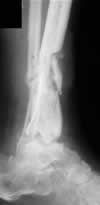
Initial x-rays of an infected tibial nonunion in a 77 year old man 27 months after his injury. He had undergone three previous surgeries at an outside facility by other physicians but had active drainage from a wound over the fracture site at the time he was referred to our office. He had severe pain, even at rest, and was confined to a wheelchair and unable to walk.
Treatment in Progress
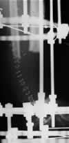
This x-ray shows the 4½ inch gap that resulted from removing all of the infected bone. The Ilizarov external fixator can also be seen in place.
Treatment in Progress
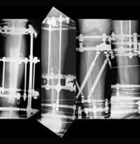
This series of x-rays taken over several weeks shows bone transport, or movement, of a segment of the tibia from near the knee toward the ankle. New bone forms as the bone moves, and the segment eventually compresses the nonunion (last picture on right) to stimulate healing.
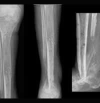
Result After Treatment by Dr. Brinker
Following treatment, these x-rays show the patient’s new bone tissue that was formed in the upper tibia (left image) and the fusion of the lower tibia with the foot (middle and right images). Stability has been restored, infection has been eliminated, the patient has occasional minimal pain and can walk without assistance.
Infected Nonunion: Tibia Case 3
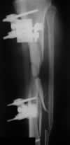
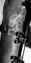
Presenting
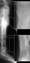
This x-ray and photograph show a 37 year old man with an infected tibial nonunion that had been treated at an outside facility by other physicians with a unilateral external fixator for four months before referral into our office. He had more than 4 inches of infected bone in the middle part of his tibia, as well as skin breakdown and drainage at the wound site.
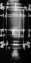
Treatment in Progress
This x-ray during treatment shows a double-level Ilizarov bone transport. The small separated segment near the knee is being transported downward, while the larger lower segment near the ankle is being transported upward. These two segments eventually made contact in the middle of the tibia, as new bone tissue formed behind each.
Result After Treatment by Dr. Brinker
These final x-rays after 9 months of treatment show a solidly healed tibia, with new bone tissue at the upper and lower ends of the tibia. The patient has no signs of infection, minimal to no pain, and can walk without assistance.
Infected Nonunion: Tibia Case 4

Presenting
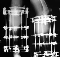
This 60 year old gentleman sustained a high energy open tibia fracture when he was injured at work. Following more than 10 surgical procedures performed at outside facilities by other physicians, the patient’s worker’s compensation nurse case manager brought the patient in for Dr. Brinker’s evaluation.
The patient was noted to have poor mechanical stability and signs and symptoms consistent with an infected tibial nonunion.
Treatment in Progress
Dr. Brinker performed serial debridements with antibiotic bead exchanges to treat the patient’s infection. This was followed by Ilizarov treatment with a double level bone transport (choo-choo train transport distal to proximal).
Result After Treatment by Dr. Brinker
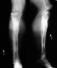
At final follow-up after Ilizarov removal the patient’s tibia is solidly healed at both transport sites and at the docking site. The patient is pain free and ambulates fully weight bearing.
Infected Nonunion: Tibia Case 5

Presenting
This 47 year old gentleman was working at a machine shop when a part came off the machine at a high velocity and impacted his left tibia. This massive blow resulted in a severe left open tibia fracture. This patient was treated at an outside facility with a spanning external fixator, serial irrigation and debridements and soft tissue coverage.
Several months later the patient presented to see Dr. Brinker with gross purulent drainage and a severely infected tibial nonunion.
Treatment in Progress
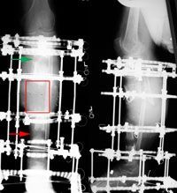
Following aggressive debridement of infected and necrotic bone, Dr. Brinker performed Ilizarov external fixation with a double level transport. The red rectangle illustrates the bony defect; the green arrow shows the proximal corticotomy site with early new bone formation; the red arrow shows the distal corticotomy site with early new bone formation.

Result After Treatment by Dr. Brinker
Final radiographs show solid healing of both corticotomy sites and the midshaft docking site. The patient is pain free, infection free, can ambulate fully weight bearing without aides, and has returned to full duty at the machine shop.
Texas Orthopedic Hospital
7401 Main St
Houston, TX
77030
Make An Appointment
Maps & Directions
Need An Expert Opinion?
Patient stories
Choose Your Affected Bone/Joint