Deformities Tibia Case
Nonunion: Tibia Case 1
Presenting
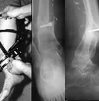
This photograph and x-rays show a 58 year old woman 14 months after an open fracture with a fixed deformity of the lower tibia following treatment at an outside facility by other physicians. The photograph shows the matching of the Ilizarov external fixator to the patient’s deformity. The patient was confined to a wheelchair and unable to position her foot to be able to bear weight effectively.
Treatment in Progress
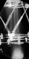
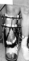
This x-ray and photograph taken during treatment show reduction of the deformity and the patient bearing weight while the Ilizarov external fixator is in place. Weightbearing often assists in stimulating bone healing.
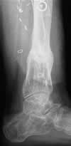
Result After Treatment by Dr. Brinker
Final lateral x-ray shows solid healing of the tibia and restoration of alignment of the limb. The patient now walks without assistance.
Deformities: Tibia Case 2
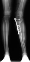
Presenting
This x-ray shows a 36 year old man 17 months after a fracture with a fixed deformity of the upper tibia following treatment at an outside facility by other physicians. He had to walk with crutches and was having back pain from his altered walking pattern due to the deformity.
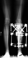
Treatment in Progress
This x-ray taken during treatment show reduction of the deformity and the patient bearing weight while the Ilizarov external fixator is in place. Weightbearing often assists in stimulating bone healing.
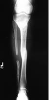
Result After Treatment by Dr. Brinker
Final x-ray shows solid healing of the tibia and restoration of alignment of the limb. The patient now walks without assistance.
Texas Orthopedic Hospital
7401 Main St
Houston, TX
77030
Make An Appointment
Maps & Directions
Need An Expert Opinion?
Patient stories
Choose Your Affected Bone/Joint