Deformities Knee
Deformities: Knee
Presentation
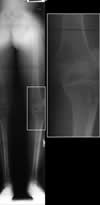
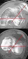
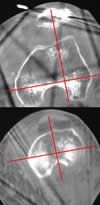
These x-rays and cross-sectional CT scans show a 43 year old man 6 months after an injury in which he was pinned under some machinery. He had an open knee dislocation, injury to the blood vessels, and disruption of all ligments in his knee. Upon referral to Dr. Brinker, he had a fixed dislocation of his kneecap and a fixed dislocation of his knee joint in a rotated position, which can be seen by the acute angle of the red lines in the right image—these lines should form a right angle. He had no motion at the knee, had constant severe pain, and was unable to bear weight on that leg.
Treatment in Progress
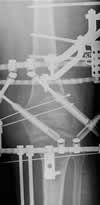
This x-ray and cross-sectional CT just before removal of the Ilizarov external fixator show restoration of kneecap and knee joint alignment; the red lines now form a normal right angle.
Result After Treatment by Dr. Brinker
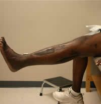
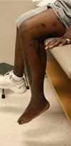
These photographs after treatment show the patient with good knee joint motion and function. He has no pain and was able to walk without assistance.
Texas Orthopedic Hospital
7401 Main St
Houston, TX
77030
Make An Appointment
Maps & Directions
Need An Expert Opinion?
Patient stories
Choose Your Affected Bone/Joint